

The 2026 Nobel Prize in Physiology or Medicine honors Karl Deisseroth, Peter Hegemann and Georg Nagel for discoveries behind optogenetics. Learn how light-gated ion channels work, what the method has revealed about brain circuits, and why experimental vision-restoration research is promising but not yet routine treatment.
Evidence classification has not been added yet. Read the full article and source card below for details.
- Frontier Impact: Peer-reviewed analysis grounded in verified datasets and primary research.
- Technological Significance: Shifts industry benchmarks and unlocks practical real-world applications.
- Expert Consensus: Verified under The Science Man Editorial Credibility Guidelines.
The 2026 Nobel Prize in Physiology or Medicine honors Karl Deisseroth, Peter Hegemann and Georg Nagel for discoveries behind optogenetics. Learn how light-gated ion channels work, what the method has revealed about brain circuits, and why experimental vision-restoration research is promising but not yet routine treatment.
The 2026 Nobel Prize in Physiology or Medicine recognizes a remarkable way of asking the brain a precise question: what happens when this particular group of nerve cells is switched on, or switched off, at this exact moment? Karl Deisseroth, Peter Hegemann and Georg Nagel share the prize for discoveries concerning light-gated ion channels and optogenetics. Their work brought together molecular biology, neuroscience and optics, giving researchers a tool to control selected cells with flashes of light and observe what follows.
Optogenetics is often described as using light to control the brain. That short description is memorable, but it can leave the wrong impression. The method does not let a researcher shine a lamp through a person’s skull and choose a thought. It is a laboratory technique that combines genetic targeting with light-sensitive proteins and carefully delivered illumination. It has transformed experiments in cells and animals, and a small number of related therapeutic approaches are now being tested in people. Those early clinical studies are promising, but experimental treatments are not the same thing as established or approved care.
This guide explains what the three laureates discovered, how optogenetics works, why the Nobel Committee considers the work important, what it has taught scientists about neural circuits, and where the medical possibilities stand today. It also separates what researchers can do now from what remains a research goal.
The 2026 Medicine Nobel Prize: who won and why?
The Nobel Assembly at Karolinska Institutet announced on 5 October 2026 that the prize would be shared equally by Karl Deisseroth, Peter Hegemann and Georg Nagel. Its stated motivation is “for their discoveries concerning light-gated ion channels and optogenetics.” In plain language, the award recognizes the discovery and development of tools that make it possible to use light to influence the electrical activity of genetically selected cells.
Hegemann and Nagel helped reveal that certain single-celled organisms contain proteins that respond to light by opening channels in their cell membranes. Deisseroth and collaborators helped establish how light-sensitive proteins could be introduced into selected mammalian neurons and used to control their activity. The contribution was cumulative: a molecular discovery in algae became a powerful experimental approach for neuroscience.
The award is not for one device or one clinical treatment. It recognizes foundational discoveries and the experimental method that grew from them. Optogenetics is now used by laboratories around the world to investigate how particular neural populations contribute to movement, sensation, learning, sleep, emotion and disease models. The method’s value lies in enabling more direct tests of cause and effect in living biological systems.
Feature portraits: Karl Deisseroth (Christopher Michel, CC BY-SA 4.0, via Wikimedia Commons); Peter Hegemann (Millencolin, CC BY-SA 4.0, via Wikimedia Commons); Georg Nagel (Millencolin, CC0, via Wikimedia Commons).
Meet the three laureates
Karl Deisseroth: connecting light-sensitive proteins with neuroscience
Karl Deisseroth is an American psychiatrist and bioengineer at Stanford University. His research brought together clinical questions about the brain with engineering approaches and molecular tools. The challenge was not simply to find a protein that responded to light; it was to make that response useful in the complex environment of a mammalian nervous system.
Researchers needed a way to put the light-responsive protein into a chosen cell population, deliver light to that population, and measure how the cells and the wider circuit responded. Deisseroth’s work helped establish and popularize this combination. Genetic instructions could be delivered so that a light-sensitive protein was made in selected neurons; optical fibers or other light-delivery arrangements could then provide timed pulses; electrophysiology and behavior could reveal the result.
This created an experimental strategy for testing whether activity in a defined group of cells is sufficient or necessary for a behavior or circuit event. The answer depends on careful controls and sound interpretation, but the approach gave neuroscience a new kind of precision. Deisseroth’s role was part of a wider collaborative history involving many laboratories, engineers, molecular biologists and neuroscientists.
Peter Hegemann: asking how algae sense light
Peter Hegemann is a German biophysicist at Humboldt University of Berlin. A central clue came from the green, single-celled alga Chlamydomonas reinhardtii. This organism swims toward or away from light, behavior that helps it find conditions useful for photosynthesis. To navigate, it must detect light and convert that information into a change in cellular activity.
Hegemann studied the alga’s light responses and helped identify the molecular machinery behind them. This line of work was fundamental research: the goal was to understand how a microorganism detects light, not to invent a neuroscience tool. Yet the biological system contained precisely the kind of switch that other researchers were looking for—a membrane protein that could respond directly to illumination.
Hegemann’s contribution shows how research driven by curiosity can later become useful in an unexpected field. The discovery did not emerge from a plan to control mammalian brains. It followed from detailed work on a tiny organism and a basic question about its biology.
Georg Nagel: identifying a light-gated ion channel
Georg Nagel is a German physiologist and biophysicist at the University of Würzburg. Working with Hegemann and colleagues, he helped characterize channelrhodopsins, proteins found in green algae that respond to light by allowing charged particles, or ions, to cross a cell membrane. Channelrhodopsin-2, usually shortened to ChR2, became a particularly useful tool.
The word “channel” matters. Many light-sensitive proteins trigger a chain of chemical steps inside a cell. A channelrhodopsin can instead form a pathway through the membrane itself. When activated by light of an appropriate wavelength, it opens and permits ions to flow. Because ion movement changes the electrical state of a cell, the protein can link a flash of light to a rapid change in cellular voltage.
Nagel’s electrophysiological and molecular work helped show how these proteins function. That understanding made it possible for other researchers to test them in new cell types. The scientific achievement was not merely noticing that algae are attracted to light; it was identifying and characterizing the molecular mechanism that could be adapted as a controllable switch.
What is optogenetics?
Optogenetics combines two broad ideas. “Genetics” refers to putting genetic instructions for a light-sensitive protein into selected cells, so those cells produce the protein. “Opto” refers to using light to activate or inhibit the protein. In a typical neuroscience experiment, researchers first choose a cell type or circuit, then use molecular targeting to make that population light-responsive. They deliver a chosen pattern of light and record the electrical, chemical or behavioral effects.
The targeting step is essential. A light pulse alone does not know which neuron matters. Scientists use promoters, viral vectors, genetic mouse lines or other molecular strategies to influence where and when the opsin is expressed. The exact approach depends on the species, tissue, research question and safety requirements. Specificity is never automatic: investigators need to verify which cells contain the protein and how much expression occurred.
After expression, light can be delivered through an optical fiber, an implanted LED, a microscope objective or another device appropriate to the experiment. The system needs enough light at the target cells without causing excessive heating or tissue damage. Researchers then measure the response. Depending on the opsin, wavelength and cell type, a pulse can increase activity, suppress it, or change the cell’s responsiveness over a longer period.
Optogenetics is a family of methods rather than a single switch. Different opsins respond to different colors, have different speeds and require different light intensities. Some are channels; others are pumps or engineered proteins that alter signaling in other ways. The tool must be matched to the experiment. A method that is ideal for rapidly activating a neuron may not be best for long-term inhibition or for a tissue that is difficult to illuminate.
How a flash of light can change a cell’s electrical activity
Every neuron maintains a difference in electrical charge between the inside and outside of its membrane. This membrane potential is shaped by ions such as sodium, potassium, chloride and calcium, as well as by pumps and channels that regulate their movement. A neuron communicates when inputs shift its voltage enough to trigger electrical signals called action potentials.
When a light-sensitive channel such as ChR2 is present in the membrane, a light pulse can open the channel. Positively charged ions flow down their electrochemical gradients, changing the membrane potential. In many commonly used settings, the result is depolarization that makes an action potential more likely. The precise result depends on the cell, its internal state, the strength and duration of light, and the opsin used.
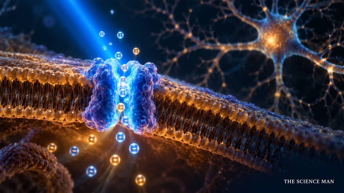
Other opsins can inhibit activity. Some move ions across the membrane in a way that makes firing less likely; some open channels that shift voltage in an inhibitory direction. Researchers can choose among several naturally occurring and engineered proteins. This gives experiments a useful vocabulary: activate a population, inhibit it, or deliver a patterned sequence and see whether timing changes the result.
Light is not a magic command. It changes the probability and timing of cellular events, and those changes occur within a living network. A neuron receives many inputs, and other cells continue to function. Turning on one population may produce no obvious behavior, may alter a circuit only in a particular context, or may lead to a measurable response. A careful experiment asks a narrow question and uses controls to distinguish the intended effect from heating, expression artifacts, light leakage or unrelated handling.
Why the method was a major advance
Before optogenetics, researchers already had ways to study neural activity. Electrical stimulation could activate tissue, but it often influenced multiple nearby cell types and fibers. Drugs could affect selected receptors, yet their effects might be slow, diffuse or difficult to reverse quickly. Lesions could show that a region mattered, but they could not reveal what happened when that region was active for a brief moment. Each method remains useful; the limitation was that it could be difficult to manipulate a defined population with both cellular specificity and millisecond-scale timing.
Optogenetics helped close that gap. A researcher could select cells by their genetic identity, deliver brief light pulses and compare activity or behavior with a control condition. This made it possible to probe circuits on the time scale at which neurons communicate. It also let researchers repeatedly switch a population on and off, which can be more informative than permanently damaging or silencing tissue.
The advance is best understood as experimental control. Observing that two brain regions are active at the same time does not establish that one causes the other or that either is responsible for a behavior. A targeted intervention can test a causal hypothesis: if this cell population is activated at this point in a task, does the outcome change? If silencing it produces the opposite effect, confidence in the interpretation can increase.
Even then, no single experiment proves an entire theory. Opsins may be expressed in unintended cells; light may spread; stimulating an artificial pattern may not mimic natural activity; and the brain may compensate. Strong conclusions come from converging evidence, replication, appropriate controls and complementary methods. Optogenetics is powerful because it adds a precise intervention to that toolkit, not because it removes the need for careful reasoning.
From algae to brain circuits: the discovery’s path
The path from algal phototaxis to modern neuroscience illustrates how basic research can travel across disciplines. A unicellular alga has a simpler organization than a mammalian brain, but it still must transform an environmental cue into a cellular response. Investigators studying that response found microbial opsins, a family of proteins that use light to influence cellular processes.
Once channelrhodopsins were characterized, researchers asked whether the proteins could function in neurons. Neurons are electrically active cells with membranes that already use ion channels to generate signals. If a light-gated channel could be expressed there, it might provide an external way to control electrical activity. Experiments in cultured cells and animal preparations tested that possibility. The tools were refined so that different populations could be targeted and light delivered with increasingly precise timing.
Turning a molecular discovery into a dependable neuroscience method required more than transferring one gene. Researchers had to solve problems in gene delivery, cell targeting, opsin expression, wavelength choice, illumination, measurement and analysis. They also had to learn what the intervention itself changed. A protein can alter membrane properties even without light, and a bright stimulus can warm tissue. Controls that account for these factors are part of the method’s development.
As the tool became more accessible, laboratories applied it to many questions. The result was a broad change in how circuits could be tested. Instead of only observing which neurons fire during an action, researchers could intervene in a selected group and ask whether that activity changes the action. That shift—from correlation toward controlled perturbation—is one reason the work has had such wide influence.
What optogenetics has helped scientists investigate
Movement and motor control
Movement depends on coordinated activity across the cortex, basal ganglia, cerebellum, spinal cord and peripheral nerves. By activating or inhibiting defined populations in animal models, researchers can test how particular pathways contribute to starting, shaping or stopping movement. The technique can help distinguish a circuit that generates a movement from one that changes its probability, speed or precision.
These experiments are relevant to conditions such as Parkinson’s disease, where specific networks behave differently, but a laboratory circuit result is not itself a treatment. A researcher may learn that altering one pathway changes a motor behavior in a mouse, yet translating that insight requires additional work on human biology, delivery, safety and meaningful clinical outcomes. Optogenetics is valuable here as a way to sharpen basic understanding before possible therapies are designed.
Sensation and perception
Touch, pain, vision and hearing each begin with specialized sensory cells and continue through several stages of neural processing. Optogenetic experiments can help test which cells carry particular signals and how downstream circuits respond. In a controlled animal study, a brief stimulus to a selected sensory pathway may reveal whether that population can drive a measurable response or whether another pathway is required.
Such findings can inform models of sensory processing and disease. They can also help researchers understand what kinds of neural activity might be useful to restore when part of a sensory system has been damaged. However, producing a response in a model organism does not mean a complex human percept can be recreated. Sensory experience depends on patterns across networks, prior learning and context, so simple claims that light can “create any sensation” overstate what the method shows.
Learning, memory and emotion
Learning changes the strength and organization of connections among neurons. Memory experiments use optogenetic tools to test whether particular cell ensembles become active during learning, whether reactivating those ensembles influences later behavior, and how context changes that response. In animal models, researchers can manipulate tagged populations and study recall, fear learning, reward and other behaviors.
Popular descriptions sometimes claim that scientists can switch memories on or off at will. That wording is too broad. Experimental manipulation may alter a measured behavior associated with a learned cue in a particular model; it does not mean that a human memory is a single file that can be edited cleanly. Memories are distributed, reconstructed and shaped by context. The method helps investigate the biology of memory, but it does not amount to precise control of a person’s thoughts or identity.
Sleep and internal states
Sleep and wakefulness involve interacting brain systems that regulate arousal, circadian timing and transitions among states. Optogenetic studies can test whether activity in a particular population is enough to promote wakefulness or a sleep phase in an animal under defined conditions. The timing of stimulation is important because the same intervention can have different effects depending on the state of the larger network.
For clinicians and patients, these studies are a source of mechanistic hypotheses, not ready-made advice. They do not justify self-treatment with light or imply that a consumer light device can reproduce an experiment. Laboratory tools use engineered cellular expression and controlled protocols that are not equivalent to ordinary exposure to colored light.
Disease models and circuit dysfunction
In models of epilepsy, psychiatric conditions and neurodegenerative disease, optogenetics can help identify circuit imbalances. For example, activating an inhibitory population or reducing activity in an overactive pathway may change seizures or a disease-related behavior in an animal model. Such work helps test mechanisms and can reveal which cells are worth studying with other techniques.
There is an important boundary between a useful model and a human diagnosis. Animal models reproduce selected features of a disease, not every experience or biological cause in a person. An optogenetic result can suggest a pathway for investigation; it does not establish that directly manipulating the same pathway would be safe or beneficial in patients. Clinical therapies must meet separate standards for evidence, delivery, durability, risks and effectiveness.
Optogenetics in the clinic: experimental vision restoration
One of the clearest attempts to translate optogenetic principles into human medicine involves the retina. Retinitis pigmentosa is a group of inherited disorders in which light-sensing photoreceptor cells progressively degenerate. In advanced disease, some inner retinal cells may remain even after rods and cones have been severely damaged. Researchers have explored whether gene delivery could make surviving cells respond to light, effectively giving them a new light-sensing role.
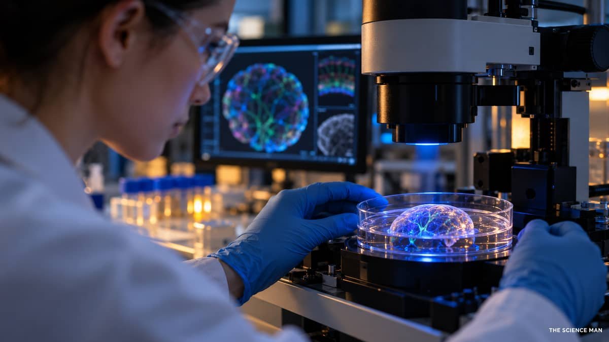
In a 2021 Nature Medicine report, a patient with advanced retinitis pigmentosa received an adeno-associated viral vector carrying instructions for the light-sensitive protein ChrimsonR. The treated eye was paired with engineered goggles that detected visual scenes and projected timed light pulses onto the retina. The report described the patient perceiving, locating, counting and touching some objects with the treated eye while wearing the goggles. It was a significant proof of concept, but it involved one patient and did not restore ordinary vision.
On 7 October 2026, investigators reported a ten-participant study in the New England Journal of Medicine. The open-label study evaluated optogenetic therapy for people with advanced retinitis pigmentosa. According to the published report and the participating institutions’ summary, several participants showed meaningful improvement in light sensitivity, and some performed better on simple object-related tasks when using the stimulation goggles. The results are early clinical evidence that the approach can produce aspects of visual function in some people.
The limitations matter as much as the result. The study was small, and participants did not regain normal high-resolution vision. The goggles and training were part of the intervention; the treated retina did not simply become equivalent to a healthy eye. The work does not show that all blindness can be reversed, or that the approach is suitable for every retinal disease. It remains a specialized investigational therapy requiring clinical studies and long-term assessment.
There are additional scientific and practical questions. Researchers must determine how durable the effect is, how consistently different people respond, what training is needed, how to improve resolution and contrast, and how to measure benefits that matter to patients. Viral gene delivery also requires careful evaluation of dose, immune response and eye-specific risks. Because each optogenetic therapy may use a different protein, vector, delivery route or device, findings from one program cannot automatically be applied to another.
As of this article’s publication date, the evidence described here should be treated as experimental clinical research, not as a broadly available or established treatment. Anyone with an inherited retinal condition should discuss current care and legitimate trial eligibility with an ophthalmologist or inherited-retinal-disease specialist. A headline about restored sight does not tell an individual patient whether a specific trial is appropriate.
Why direct optogenetic control of the human brain is difficult
Most optogenetic neuroscience experiments require both a genetic step and a light-delivery step. In a laboratory animal, researchers can use a carefully designed vector or genetic line to make selected cells express an opsin, then deliver light through an optical fiber or implanted source. Applying the same arrangement throughout a person’s brain would raise major technical, medical and ethical questions.
Light does not travel deeply through opaque tissue in a focused, controllable way. Delivering light to a deep brain target could require an implant, which is invasive and would have to remain safe over time. Gene delivery would need to reach the right cells with adequate specificity and without harmful effects. A therapy would also need a reliable way to control the stimulation, monitor its effects and manage variability among patients.
For these reasons, the clinical applications of optogenetics are not simply a matter of scaling up a mouse experiment. The retina offers a more accessible setting because it is part of the eye and can be reached with specialized approaches, though ocular gene therapy still has substantial risks and limitations. Research on noninvasive stimulation or other therapeutic technologies may be inspired by circuit discoveries, but those approaches are not optogenetics unless they use light-sensitive proteins in target cells.
Benefits and limitations of the method
Strengths
- Cell-type targeting: genetic strategies can focus an intervention on selected populations, subject to the specificity of the targeting system.
- Fast timing: many opsins respond on a time scale useful for studying rapid neural signaling.
- Reversible experiments: researchers can deliver pulses, stop them and compare conditions in the same preparation.
- Direct causal tests: carefully controlled activation or inhibition can test whether a population contributes to a circuit function.
- Flexible design: a growing set of opsins and delivery systems lets scientists adapt the tool to a question.
Limitations
- Expression is not perfect: genes may be expressed in cells outside the intended population or at levels that change cell behavior.
- Light delivery is challenging: tissue scatters and absorbs light, which limits precision and depth and can cause heating at high intensities.
- Artificial patterns may not be natural: a synchronized light pulse can drive cells differently from their normal input patterns.
- Biology varies: cell type, species, developmental stage and disease state can change the response.
- Translation is slow: safety, delivery, durability and clinical benefit need to be established independently for every therapy.
These limits are not reasons to dismiss optogenetics. They are reasons to design experiments carefully and report their scope honestly. Every powerful method has conditions under which it works well and assumptions that can fail. The best studies pair optogenetic manipulation with measurements such as electrophysiology, imaging, anatomy and behavior, and they include light-only, vector-only and other appropriate control conditions.
How optogenetics compares with other neuroscience tools
Electrical stimulation changes activity by applying current through an electrode. It can be useful clinically and experimentally, but the current may affect multiple cell types near the electrode. Optogenetic targeting can be more selective by cell identity, although it requires genetic expression and light access. The two techniques answer different questions and may complement one another.
Pharmacology changes receptor or enzyme activity using a chemical. Drugs can reach many cells and may have effects lasting minutes, hours or longer. This broad reach can be an advantage when researchers want to study a whole pathway or when a medicine needs to act throughout a body. Optogenetics can offer faster control in a defined population, but it is more technically demanding and is not a replacement for medication development.
Functional imaging measures signals associated with neural activity across many cells, often without directly manipulating them. Imaging can reveal where and when activity changes but usually provides an observational signal. Optogenetics can provide an intervention; combining stimulation with imaging can help link a circuit change to an outcome. The combination still requires careful controls because optical stimulation and optical measurement can interfere with each other.
Lesions and genetic knockouts can show what happens when a structure or gene is absent. They are informative for necessity but may trigger adaptation over time. Optogenetics can turn activity up or down on a chosen time scale, helping distinguish immediate circuit effects from long-term compensation. Each method reveals a different layer of biology. Strong neuroscience uses multiple approaches rather than crowning one as universally best.
Ethics, animal welfare and responsible reporting
Much of optogenetics’ impact has come from animal research. Such work must be reviewed under institutional and national rules, designed to use the fewest animals consistent with a meaningful result, and carried out with appropriate welfare protections. The method can reduce some kinds of invasive manipulation, but genetic modification, surgery, implants and repeated testing still require careful ethical oversight.
Human applications bring additional concerns. Gene delivery may be difficult to reverse, and long-term outcomes need study. Researchers must explain uncertainty clearly, obtain informed consent and avoid overstating the likelihood of benefit. In conditions with limited treatment options, people may be especially vulnerable to claims that an experimental intervention will restore sight or cure disease. Accurate communication distinguishes a trial result from a proven therapy.
The phrase “controlling the brain with light” can make for a striking headline, but it can also imply control over a whole person. The actual intervention is narrower: selected cells that have been engineered to express a protein respond to a defined stimulus. Cells influence circuits, circuits influence behavior, and the resulting behavior depends on many conditions. Responsible reporting preserves that distinction and avoids portraying patients as having their thoughts or personalities externally directed.
What could come next?
On the research side, scientists continue to develop proteins that respond to different wavelengths, operate at lower light levels, switch faster or allow finer control. Better targeting systems and less invasive illumination could extend what experiments can address. Miniaturized devices, improved imaging and computational analysis may help measure more cells and connect cellular activity to behavior with greater precision.

For medicine, progress will depend on more than improved opsins. Researchers need reliable gene delivery, predictable expression, safe devices, clear measures of meaningful benefit and evidence from larger, appropriately designed trials. For vision research, future studies will need to learn which patients are most likely to benefit and whether training and device improvements can support useful everyday function. Each step should be judged against outcomes that patients value, not only laboratory signals.
There may be indirect clinical effects even when optogenetics itself is not used as a treatment. By showing how specific circuits contribute to a disease-related behavior, the method can help identify targets for drugs, electrical stimulation, rehabilitation or other interventions. A research tool can influence medicine by improving understanding, even if it never becomes a therapy used directly in most patients.
Predictions should remain modest. A discovery can open a route without guaranteeing that the route leads to a safe or effective treatment. The history of biomedical science contains many promising mechanisms that did not translate as expected, as well as basic discoveries whose practical value emerged decades later. The most defensible claim is that optogenetics has changed the questions scientists can test and is now inspiring carefully monitored clinical research.
The last ten Nobel Prizes in Physiology or Medicine
The 2026 award sits within a decade of discoveries across many scales of biology, from the molecular clock and immune regulation to RNA and neural control. The table below lists the ten most recent awards, from 2017 through 2026.

| Year | Laureates | Recognized discovery or contribution |
|---|---|---|
| 2026 | Karl Deisseroth, Peter Hegemann, Georg Nagel | Discoveries concerning light-gated ion channels and optogenetics. |
| 2025 | Mary E. Brunkow, Fred Ramsdell, Shimon Sakaguchi | Discoveries concerning peripheral immune tolerance. |
| 2024 | Victor Ambros, Gary Ruvkun | Discovery of microRNA and its role in post-transcriptional gene regulation. |
| 2023 | Katalin Karikó, Drew Weissman | Discoveries concerning nucleoside base modifications that enabled effective mRNA vaccines against COVID-19. |
| 2022 | Svante Pääbo | Discoveries concerning extinct hominin genomes and human evolution. |
| 2021 | David Julius, Ardem Patapoutian | Discovery of receptors for temperature and touch. |
| 2020 | Harvey J. Alter, Michael Houghton, Charles M. Rice | Discovery of the hepatitis C virus. |
| 2019 | William G. Kaelin Jr, Peter J. Ratcliffe, Gregg L. Semenza | Discoveries of how cells sense and adapt to oxygen availability. |
| 2018 | James P. Allison, Tasuku Honjo | Discovery of cancer therapy by inhibition of negative immune regulation. |
| 2017 | Jeffrey C. Hall, Michael Rosbash, Michael W. Young | Discoveries of molecular mechanisms controlling the circadian rhythm. |
These awards do not form a ranking of medical importance. Nobel Prizes recognize particular contributions selected by the relevant committee, and each entry reflects a long history of work by many scientists. The 2026 prize highlights one of the modern era’s major experimental methods: a way to intervene in living neural systems with much finer control over cell type and timing than many earlier approaches allowed.
Frequently asked questions
Who won the 2026 Nobel Prize in Physiology or Medicine?
Karl Deisseroth, Peter Hegemann and Georg Nagel share the 2026 prize for discoveries concerning light-gated ion channels and optogenetics. The prize was announced on 5 October 2026.
What does optogenetics mean?
Optogenetics combines genetic methods that make selected cells produce light-sensitive proteins with light used to activate or inhibit those proteins. Scientists use the approach to study cellular activity and neural circuits.
Can optogenetics control a person’s thoughts?
No. That popular shorthand is misleading. Experimental optogenetics acts on selected cells that have been engineered to express light-sensitive proteins, usually in controlled laboratory settings. It does not provide general access to thoughts, memories or personality.
Can optogenetics cure blindness?
There is no general cure for blindness from optogenetics. Clinical studies in advanced retinitis pigmentosa have reported partial aspects of visual function for some participants using gene therapy together with specialized stimulation goggles. These approaches remain investigational and do not restore ordinary high-resolution vision.
Is optogenetic vision restoration approved treatment?
The studies described here are clinical research. A trial result should not be read as proof that a treatment is approved or available for routine care. Patients should ask their own eye-care specialist about established options and legitimate clinical trials.
Why are light-sensitive proteins found in algae useful for studying neurons?
Some algal proteins respond directly to light by opening an ion channel or moving ions across a membrane. Since neurons use ion movement to change their electrical activity, researchers can use these proteins as tools to influence selected cells with light.
Does optogenetics require surgery?
Many brain experiments in animals involve gene delivery and an implanted or fiber-based light source. The details vary by experiment. Retinal clinical approaches use a different configuration involving gene delivery to the eye and external stimulation goggles. These are specialized medical research procedures, not consumer technologies.
What is the difference between optogenetics and ordinary light therapy?
Optogenetics requires cells to express a light-sensitive protein and uses a controlled light stimulus to act on that protein. Ordinary light exposure does not add such proteins to cells and should not be assumed to reproduce optogenetic effects.
Bottom line
The 2026 Nobel Prize honors a chain of discoveries that began with the biology of light-sensing algae and grew into a method for investigating neural circuits. Hegemann and Nagel helped uncover light-gated ion channels; Deisseroth helped bring these molecular switches into the neuroscience toolkit. Optogenetics allows researchers to ask more precise causal questions about how selected cells contribute to a living system.
The method has reshaped research in neuroscience and produced early human studies in vision restoration. Those studies are important first steps, not a universal cure or a routine treatment. The lasting achievement is both conceptual and practical: by connecting genes, proteins, light and behavior, optogenetics has given science a new way to test how biological circuits work—and a carefully bounded starting point for exploring how they might one day be helped.
Sources and further reading
- Nobel Prize: 2026 Physiology or Medicine — award summary
- Nobel Prize: popular information on the 2026 award
- Nobel Prize: advanced information on light-gated ion channels and optogenetics
- Karl Deisseroth — Nobel facts; Peter Hegemann — Nobel facts; Georg Nagel — Nobel facts
- Sahel et al., “Partial recovery of visual function in a blind patient after optogenetic therapy,” Nature Medicine (2021), doi:10.1038/s41591-021-01351-4.
- Sahel et al., “Optogenetic Therapy for Restoring Aspects of Visual Function,” New England Journal of Medicine (2026), doi:10.1056/NEJMoa2602215.
- University of Basel: summary of the 2026 visual-function study.
- Nobel Prize: Physiology or Medicine laureates by year.
Portrait credits: Karl Deisseroth photograph by Christopher Michel, CC BY-SA 4.0; Peter Hegemann photograph by Millencolin, CC BY-SA 4.0; Georg Nagel photograph by Millencolin, CC0. Images via Wikimedia Commons. The featured collage identifies each laureate and retains these credits.

digitalwebman@gmail.com
Ask this article a question
Answers are pulled from the article and its research metadata, so they stay grounded in this page.
